Four technologies.One clear picture.
Every method answers a different question. MRI shows structure, blood shows how your body is working, CT and ultrasound show your heart and arteries in detail. We can recommend the correct technology for the correct concern.

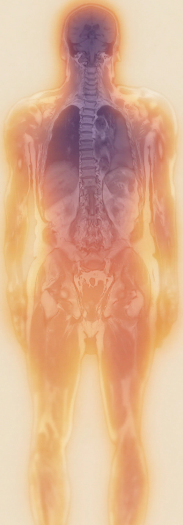
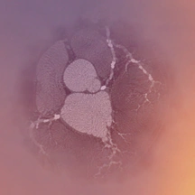
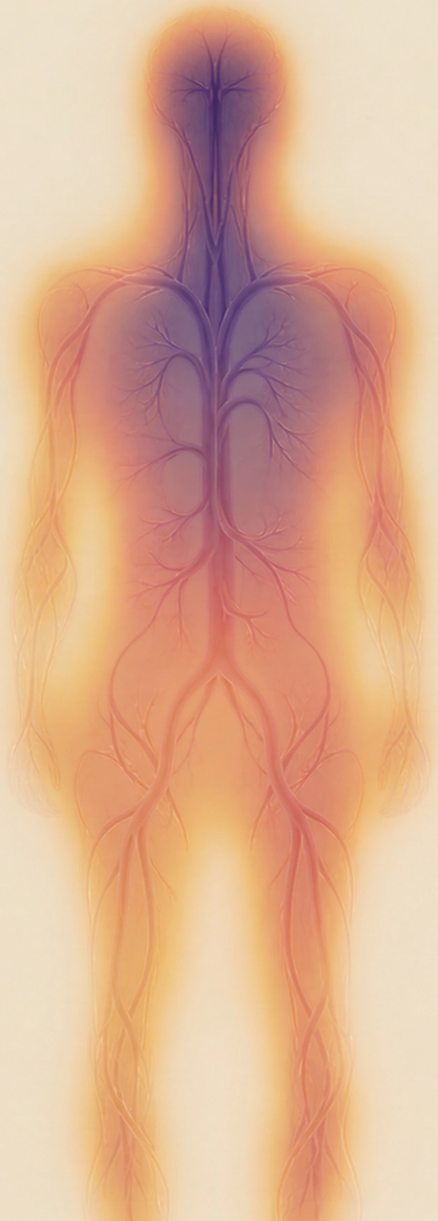

What each technology shows Four tools, examined for 500+ possible findings
Full-body MRI
Brain, organs, spine and major vessels in one session. Also the basis for body composition and breast MRI.
Blood analysis
How your heart and vessels, metabolism, liver, kidneys and thyroid are working.
Heart CT
Your coronary arteries in detail: a calcium score for calcified plaque, and CT angiography for soft plaque and narrowing.
Artery ultrasound
Plaque in the neck and groin arteries is a sign linked to a higher risk of stroke and heart attack.⁸
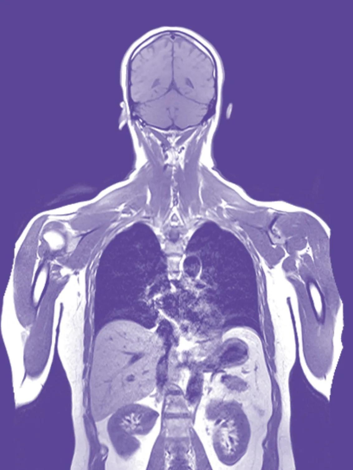
Full-body MRIA look inside, without radiation
Our protocol covers your body from head to mid-thigh. Up to 12 different tissue contrasts, including whole-body diffusion-weighted imaging (DWI), give the radiologist a detailed view of brain, organs, spine and major blood vessels. MRI works with a magnetic field and radio waves, not X-rays, and our standard protocol needs no contrast agent.
- Brain and spine
- Organs of the chest, abdomen and pelvis, such as liver, kidneys and pancreas
- Major blood vessels, including the aorta
- You feel fine and want to know where you stand
- Something runs in your family and you’d rather look than wonder
- You want a baseline to compare against in a few years
Bowel cancer, checked with a colonoscopy. Breast cancer, checked with a mammogram, which picks up tiny calcium deposits. Lung cancer, checked with a low-dose CT. Prostate cancer, checked with a PSA test and a prostate examination. The scan ends at mid-thigh and isn’t designed for detailed images of joints, such as knee or ankle injuries. None of this replaces the screening your doctor recommends.
Tell us when you book. We check what’s possible and help you feel at ease.

Blood analysisHow your body is working right now
The Holistic Health panel included with every Full Body check covers 65 biomarkers across heart and vessels, blood sugar and insulin, liver, kidneys, thyroid, iron and inflammation. Imaging shows structure, blood shows function. Read together, your blood values can help your doctor put MRI findings into context and tell harmless findings apart from those that deserve a closer look. That’s why every Full Body MRI check includes both.
Your blood values can show early signs of conditions that often cause no symptoms at first. Long-term blood sugar (HbA1c) can point to early signs of diabetes, and liver values such as ALT and GGT can indicate a fatty liver. Cholesterol, triglycerides and the inflammation marker hsCRP help estimate your risk of heart disease and stroke. Creatinine and eGFR show how well your kidneys filter, TSH shows how your thyroid is working, and your blood count can reveal unusual changes in your blood cells that deserve a closer look.
The panel spans your blood count, cholesterol and ApoB, blood sugar, HbA1c and insulin, liver and kidney values, thyroid, iron status, vitamin D and markers of inflammation such as hsCRP. Your doctor explains what each value means for you.
More about biomarkers- You often feel low on energy and want to know whether your blood values play a part
- Diabetes or high cholesterol runs in your family and you want to know where you stand
- You’re changing how you eat or train and want a baseline to measure your progress against
- Your last blood test was years ago and you want an up-to-date picture
Blood values describe how your body is working today, not a diagnosis on their own. Your doctor reads them together with your imaging and your history, and tells you which values, if any, deserve a follow-up.
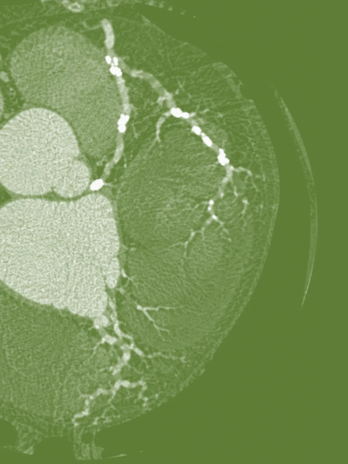
Heart CTA detailed look at your coronary arteries
A short, low-dose CT of the heart, without contrast agent. It measures calcified plaque in each coronary artery and adds it up to one number, the Agatston score. A score of zero means no calcified plaque was found.
Heart CT shows how much plaque has built up in your coronary arteries. The calcium score measures the calcified part, and the European Society of Cardiology lists it as a tool that can refine a cardiovascular risk estimate, especially when your risk sits close to a decision threshold.3 Soft plaque and narrowing don’t show up in the calcium score. For those, a coronary CT angiography with contrast agent gives a detailed view, and our medical team offers it when it’s appropriate for you.
- Heart disease runs in your family
- Your cholesterol or blood pressure is borderline and you want more to go on
- You want a first calcium score to compare against in a few years
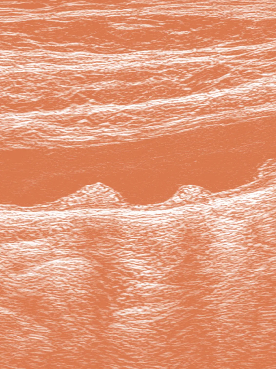
Artery ultrasoundPlaque, measured
A radiation-free ultrasound of the carotid arteries in your neck and the femoral arteries in your groin. It shows whether plaque has built up in the vessel wall, measures how much, and uses Doppler to see how your blood flows past it.
Ultrasound shows whether plaque has already formed in your arteries, something blood values alone can’t tell you. It’s more common than most people think: in the PESA study of 4,184 people aged 40 to 54 without symptoms, plaque was found in the groin arteries of 44% and in the neck arteries of 31%.5 And it matters: in the Tromsø Study, which followed more than 10,000 adults in Norway, people with larger amounts of plaque in their neck arteries had about a 65% higher risk of a first stroke or heart attack than people with none.8 That’s why European guidelines on cholesterol treatment count plaque on ultrasound as a sign of higher risk of heart attack and stroke, which can change how your cholesterol is managed.6
- Your cholesterol or ApoB is raised and you want to see whether it has left a trace
- You want a number you can measure again later
No radiation, no contrast agent. About 30 minutes, including a short walk from the MRI area to the ultrasound room.
Aeon technology
Which technology answers which question
Imaging and blood are read side by side. A finding in one can be put into context by the other, and your report brings them together.
| Main view, Partial view | MRI | Blood | Heart CT | Artery ultrasound |
|---|---|---|---|---|
| Brain and spineBrain and spine | ||||
| Organs, structureOrgans, structure | ||||
| Organ functionOrgan function | ||||
| Coronary arteriesCoronary arteries | Risk markers | |||
| Neck and groin arteriesNeck and groin arteries | ||||
| Breast tissueBreast tissue | Breast MRI (add-on) | |||
| Fat and muscle distributionFat and muscle distribution | Body composition (Full Body Plus and Premium) | |||
| Metabolism and inflammationMetabolism and inflammation | ||||
| RadiationRadiation | None | None | Low dose | None |
| Contrast agentContrast agent | Only for breast MRI | No | Only for CT angiography | No |
All your resultsIn one place
Your portal
Reports and blood values from every technology land in your Aeon portal, stored encrypted in Switzerland and available any time.
Over time
Compare results across check-ups and see what changed.
People behind the technology
Radiologists, physicians and MRI physicists, working together in Switzerland. They set the protocols, review the results and decide which technology fits which question.
Customer stories
Read more
“I had no symptoms... we discovered a small tumour... otherwise I would have waited years.”
“I had no symptoms... we discovered a small tumour... otherwise I would have waited years.”
Maria-Isabel, 50. Maria’s preventive check-up revealed a breast tumour in its earliest stage. Early detection enabled minimally invasive treatment, today Maria made a full recovery.

My sister didn’t have the chance to detect her condition through a preventive check-up....
Cathrine, 57. Cathy’s sister was diagnosed with pancreatic cancer, and it changed how she thinks about “waiting.” Too often, people are told to come back when symptoms show up.

I want to make sure that my body is fit.
Gianmaria, 36. Had a full-body scan to set his baseline and now works on his health with the Aeon intervention plan.
Frequently Asked
A whole-body MRI ("magnetic resonance imaging") scan is an imaging procedure that uses a strong magnetic field and radio waves to create high-quality images of the entire body in a single examination. This includes body parts such as head and brain, neck, chest, abdomen and pelvis, spine, joints, blood vessels, and soft tissues (muscles, tendons, and ligaments).
A whole-body scan can help detect a variety of diseases and abnormalities and derive appropriate treatment recommendations. These include tumor diseases and metastases, inflammations, infections, injuries, vascular changes, aneurysms and neurological diseases (e.g., Alzheimer's and dementia, Parkinson's, multiple sclerosis). More information can be found in our blog article "MRI 101".
A recent study found that MRI screening in adults who did not have any disease symptoms led to clinically relevant findings in about one-third of patients. Of these findings, 2.6% were confirmed by pathology as invasive cancers. Whole-body MRI is a valuable tool for early cancer detection in individuals with hereditary cancer syndromes, significantly improving their chances of long-term health and survival.
Before an MRI examination, you should inform the radiology staff about implants (breast implants, metal implants), pacemakers, medications, allergies, or pregnancy. You should also remove all metal objects and jewelry. If you have claustrophobia, be sure to let them know beforehand.
MRI, X-ray and CT are based on different technologies and are therefore suitable for different medical applications. X-rays are used to produce two-dimensional images that are particularly good at showing bone structure. Computed tomography (CT) combines X-rays with a rotating camera to produce detailed cross-sectional images, which uses more radiation but provides a more precise view of complex tissues. In contrast, magnetic resonance imaging (MRI) uses magnetic fields and radio waves to produce high-resolution images of soft tissue without radiation exposure, but with longer examination times.
A whole-body MRI as a preventive examination enables the early detection of diseases, which can lead to more effective and less invasive treatments. It offers comprehensive screening and can reveal life-changing, and thus life-saving results. It is a long-term investment in your health that saves you many other examinations.
The following values are analyzed in our Starter and Plus packages: Total cholesterol, HDL cholesterol, LDL cholesterol, non-HDL-cholesterol, Triglycerides, High-sensitivity CRP, Lipoprotein (a), Apolipoprotein B, Folic acid (folate) in serum, Vitamin B12, Creatinine, eGFR, Alanine aminotransferase (ALAT), GGT: Gamma-glutamyl transferase, TSH, Leukocytes (white blood cells), Erythrocytes (red blood cells), Hemoglobin, Hematocrit, Thrombocytes (platelets), MCV, MCH, MCHC, RDW-CV, HbA1c (NGSP), HbA1c (IFCC)
Any more questions?
You can contact our team any time and we’ll be happy to answer any of your questions.

 Contact us
Get Started
Clarity about your health.Plan your check‑up.
Book your check-up and add what makes sense for you.
Sources
- Linge J et al. Body composition profiling in the UK Biobank Imaging Study. Obesity 2018. ukbiobank.ac.uk
- Mann RM et al. Breast cancer screening in women with extremely dense breasts: recommendations of the European Society of Breast Imaging (EUSOBI). Eur Radiol 2022;32:4036–4045. PMC9122856
- Visseren FLJ et al. 2021 ESC Guidelines on cardiovascular disease prevention in clinical practice. Eur Heart J 2021;42:3227–3337. ESC summary
- American College of Radiology / RSNA. Radiation dose to adults from common imaging examinations (dose reference card). acr.org
- Fernández-Friera L et al. Prevalence, vascular distribution, and multiterritorial extent of subclinical atherosclerosis in a middle-aged cohort: the PESA study. Circulation 2015;131:2104–2113. LSHTM
- Mach F et al. 2019 ESC/EAS Guidelines for the management of dyslipidaemias. Eur Heart J 2020;41:111–188. Recommendation summarised in Swiss Medical Weekly. smw.ch
- Kronenberg F et al. Lipoprotein(a) in atherosclerotic cardiovascular disease and aortic stenosis: a European Atherosclerosis Society consensus statement. Eur Heart J 2022;43:3925–3946. review
- Eltoft A et al. Joint effect of carotid plaque and C-reactive protein on first-ever ischemic stroke and myocardial infarction? The Tromsø Study. J Am Heart Assoc 2018;7(11):e008951. ahajournals.org
- Federal Office of Public Health (FOPH). Umweltradioaktivität und Strahlendosen in der Schweiz 2024 (annual report). bag.admin.ch