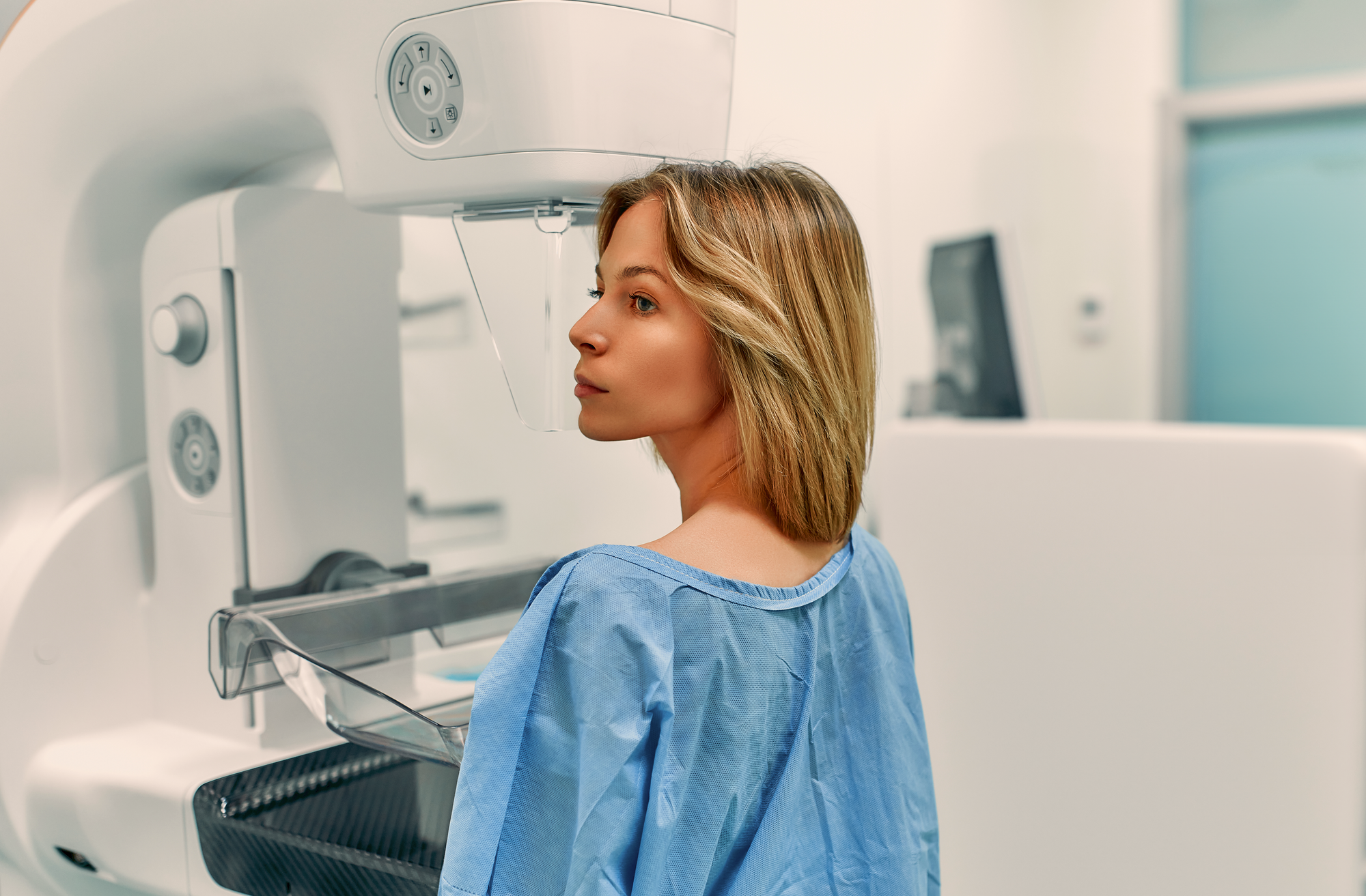
Worry about radiation is one of the most common reasons people put off a whole-body MRI. Here's the physics behind why that worry doesn't apply to MRI, and how its dose compares with a CT scan or an X-ray.
Does an MRI use radiation at all?
The short answer is no, not in the sense that matters. An MRI scanner builds its images from a strong magnetic field and radio waves, not from X-rays. Your body is mostly water, and water is full of hydrogen atoms. Put a person inside a powerful magnet and those hydrogen nuclei line up with the field. A pulse of radio waves then knocks them briefly out of alignment, and as they settle back, they release a faint signal. The scanner picks that signal up and turns it into the cross-sectional images a radiologist reads.
None of that involves ionising radiation, the kind of radiation that can damage DNA and is linked to a small rise in cancer risk over a lifetime. It's an entirely different physical process from the one a CT scanner or an X-ray machine uses, both of which fire X-ray photons through the body and measure what comes out the other side. For the fuller rundown of how MRI and CT differ beyond radiation, our comparison of the two methods goes through it step by step.
An MRI builds its images from magnetism and radio waves. There's no X-ray tube inside the machine, so there's no radiation dose to measure, no matter how the scan is set up.
How does that compare with CT and X-ray doses?
Radiation dose from medical imaging is measured in millisieverts (mSv), and the amount varies a lot by examination. A real comparison needs real numbers rather than a vague sense of "more" or "less", so here's what the research on medical radiation dose actually reports.
| Examination | Typical effective dose |
|---|---|
| MRI, any body region | None -- no ionising radiation involved |
| Low-dose CT protocol (e.g. for suspected kidney stones) | About 1.5 mSv |
| CT of the chest, abdomen or pelvis | About 10 mSv |
| Multiphase CT of the abdomen and pelvis (a higher-dose protocol, sometimes used for cancer staging) | About 31 mSv |
How these doses compare
Typical effective doses, in millisieverts (mSv). The axis breaks after 10 mSv to fit in a much higher-dose example.
To put those numbers next to something familiar: everyone absorbs some radiation just by living on Earth. Cosmic rays, radon in buildings and naturally radioactive elements in rock and soil add up to roughly 3 mSv a year on average worldwide. In Switzerland, where radon levels in parts of the Alps and the Jura push the figure up, the average is closer to 6 mSv a year, and medical imaging adds a further 1.7 mSv a year to that on average. Flying adds a little too: a round-trip flight between, say, Zurich and New York exposes you to about 0.1 mSv of cosmic radiation, which is close to the margin of error on the other numbers here. A single CT of the chest, abdomen or pelvis, in other words, delivers roughly as much as one to three years of that everyday background exposure, in one sitting, and the higher-dose, multiphase CT protocol sometimes used for cancer staging delivers several times that again. An MRI adds none of it, whatever body region is being scanned and however many scans you have.
Is that level of radiation actually something to worry about?
Radiology has spent decades studying exactly this question, because CT and X-ray are genuinely useful tools, and the goal isn't to avoid them but to use them where they add real value and keep the dose as low as the job allows. That's the thinking behind initiatives such as ALARA ("as low as reasonably achievable"), and Image Gently and Image Wisely, which focus on cutting unnecessary dose for children and adults.
The numbers researchers work with put this in perspective. Average background radiation of around 3 mSv a year isn't treated as a significant cancer risk on its own. At an effective dose of 10 mSv, roughly what a CT of the chest, abdomen or pelvis delivers, the lifetime cancer-risk model used in US radiation-protection guidance (BEIR VII) estimates about one extra case of cancer for every 1,000 people exposed at that level. That number only means something once you have the baseline it's added to: in the United States, around 42% of people develop cancer at some point in their life for reasons that have nothing to do with any scan, so researchers are describing a small increase on top of that already-substantial lifetime chance, not the total risk from the scan itself. Using the same model, the higher-dose, multiphase CT sometimes used for cancer staging, around 31 mSv, works out to roughly 3 extra cases per 1,000 people: a larger number, and part of why that protocol is reserved for situations where the diagnostic benefit clearly earns it. It's a real number either way, which is exactly why radiologists keep dose as low as the diagnosis allows, but it's a small one next to the reason the scan was ordered in the first place.
None of that applies to MRI, because the variable itself, radiation dose, simply isn't there. A CT scan carries a dose that adds up if it's repeated often; an MRI doesn't carry one at all.
If MRI carries no radiation, is it risk-free?
No, and it's worth being accurate about that rather than overselling it. An MRI's side effects, where they happen, just have nothing to do with radiation.
The scanner itself is a narrow tube, and some people find that uncomfortable, or find the knocking noise from the magnetic coils switching on and off unpleasant. We've covered how to prepare for that, including what actually helps with claustrophobia during an MRI, separately. At Aeon, you can listen to your own music during the scan, and you're in contact with the team throughout.
If a contrast agent is used, it's gadolinium-based rather than the iodine-based agents used in CT, and reactions are uncommon: occasional itching, a rash or nausea, and, rarely, a more serious allergic reaction. Our piece on contrast agents in radiology covers when they're actually needed and what the side effects look like. Aeon's own full-body scans are set up so that a contrast agent generally isn't needed in the first place.
MRI also isn't suitable for everyone. A strong magnetic field can interact with certain implants, such as some pacemakers or metal fragments in the eye, so anyone with implants or embedded metal should flag it before the appointment.
What this means if you're considering a full-body scan
Put the two differences together and the practical takeaway is straightforward. A CT scan is fast and very good at certain jobs, but it carries a radiation dose that has to be weighed against the benefit each time, which is part of why repeat CT isn't something radiologists hand out casually. An MRI removes that particular trade-off entirely. It takes longer, and it has its own limits, it isn't the best choice for assessing bone detail, for instance, but radiation dose simply isn't one of the things you need to weigh up.
That's one reason a whole-body MRI check-up works well as something people repeat over the years rather than a one-off: there's no cumulative dose to track, whatever the interval between scans.
The short version
Conclusion
Radiation is a fair question to ask about any kind of medical imaging, and it has a genuinely different answer for MRI than it does for CT or X-ray. An MRI uses magnetism and radio waves to build its images, so there's no dose to add up, however often it's repeated. That's one of the reasons a whole-body MRI makes sense as a preventive check-up you can return to, rather than a single test.
Frequently asked questions
Does an MRI use radiation in any form?
No. An MRI scanner uses a magnetic field and radio waves to build its images, not X-rays, so no ionising radiation is involved at any point in the scan.
Is it safe to have an MRI shortly after a CT scan?
Yes. Because an MRI adds no radiation dose of its own, there's nothing to add up between the two examinations. Timing only matters for the CT's own dose, not for the MRI that follows it.
Can MRI scans be repeated more often than CT scans?
In terms of radiation, yes, there's no dose-based limit on how often you can have an MRI. What limits repeat imaging in practice is cost and scanner availability, and, for CT, the cumulative radiation dose, which isn't a factor with MRI.
Is the magnetic field used in MRI itself harmful?
No published evidence links the magnetic fields and radio waves used in routine MRI to the kind of tissue damage ionising radiation can cause. The main safety consideration is metal: certain implants and metal fragments can interact with the magnet, which is why you're asked about them before every scan.
Does the contrast agent sometimes used in MRI carry a radiation risk?
No. Gadolinium-based contrast agents used in some MRIs are unrelated to radiation; the rare side effects are allergic-type reactions, not radiation exposure. Aeon's own full-body scans are set up so a contrast agent generally isn't needed.
Sources
- Jones JGA, Mills CN, Mogensen MA, Lee CI. Radiation Dose from Medical Imaging: A Primer for Emergency Physicians. West J Emerg Med. 2012;13(2):202-210. PMID: 22900113. Supports the ~3 mSv/year global background-radiation figure and the "not a significant cancer risk" framing, the ~10 mSv dose for a CT of the chest, abdomen or pelvis, the BEIR VII estimate of roughly one additional cancer per 1,000 people at that dose, the ~1.5 mSv low-dose CT protocol figure, and the ~42% US lifetime cancer baseline used for context.
- Smith-Bindman R, Lipson J, Marcus R, et al. Radiation Dose Associated With Common Computed Tomography Examinations and the Associated Lifetime Attributable Risk of Cancer. Arch Intern Med. 2009;169(22):2078-2086. PMID: 20008690. Supports the ~31 mSv figure for a multiphase CT of the abdomen and pelvis, the higher-dose, multiphase protocol sometimes used for cancer staging.
- Federal Office of Public Health (BAG), Switzerland. Radiation exposure of the Swiss population (German-language page). Supports the ~6 mSv/year average background radiation in Switzerland and the ~1.7 mSv/year average contribution from medical imaging (2023 survey figure).
- U.S. Federal Aviation Administration. Advisory Circular 120-52, Radiation Exposure of Air Carrier Crewmembers (reproduced in full at the link; original is a U.S. government publication). Table 1 gives a one-way New York-London dose of about 0.05 mSv, the basis for the ~0.1 mSv round-trip transatlantic flight figure.