Learn the difference between MRI and CT, and which examination is best suited for different medical conditions.
Most people have had at least one X-ray in their lifetime, such as for a broken bone or at the dentist. However, a Magnetic Resonance Imaging (MRI) or a Computed Tomography (CT) scan is far less common. This often leads to uncertainty about what these terms actually mean, what these examinations are used for, and how they differ.
Our article answers these questions and offers our recommendation at the end regarding which method is best suited for diagnosing specific conditions.
What Are MRI and CT? – Similarities
MRI and CT are both imaging techniques used in radiology to diagnose diseases and injuries. Both produce cross-sectional images of the inside of your body. In both cases, a contrast agent may be used to better identify certain conditions. However, at Aeon, we avoid using contrast agents to eliminate the risk of side effects. Our MRI sequences are optimized to the point where no contrast agent is necessary.
Additionally, in both examinations, you should remain as still as possible since movement can disrupt imaging and reduce the quality of the results.
How Do MRI and CT Differ?
The two biggest differences between MRI and CT concern how they work and the associated radiation exposure for patients.
A CT scan uses X-rays to visualize the body’s interior, while MRI images are created using magnetic fields and radio waves. This means that, unlike a CT scan, there is no radiation exposure with an MRI. Below, we will go into more detail about each difference.
Procedure & Functionality
MRI
An MRI scan is a simple and painless process that typically takes between 15-60 minutes, depending on the area of the body being examined.
Since MRI uses strong magnetic fields, any metal (e.g., jewelry, piercings, etc.) must be removed before the scan. If you have any metallic implants (e.g., a pacemaker), please inform the examination team during the consultation.
During the scan, you lie on the examination table inside a tube called a scanner. While the scanner captures cross-sectional images of your body’s interior, you will hear knocking noises, caused by the gradient coils constantly turning on and off.
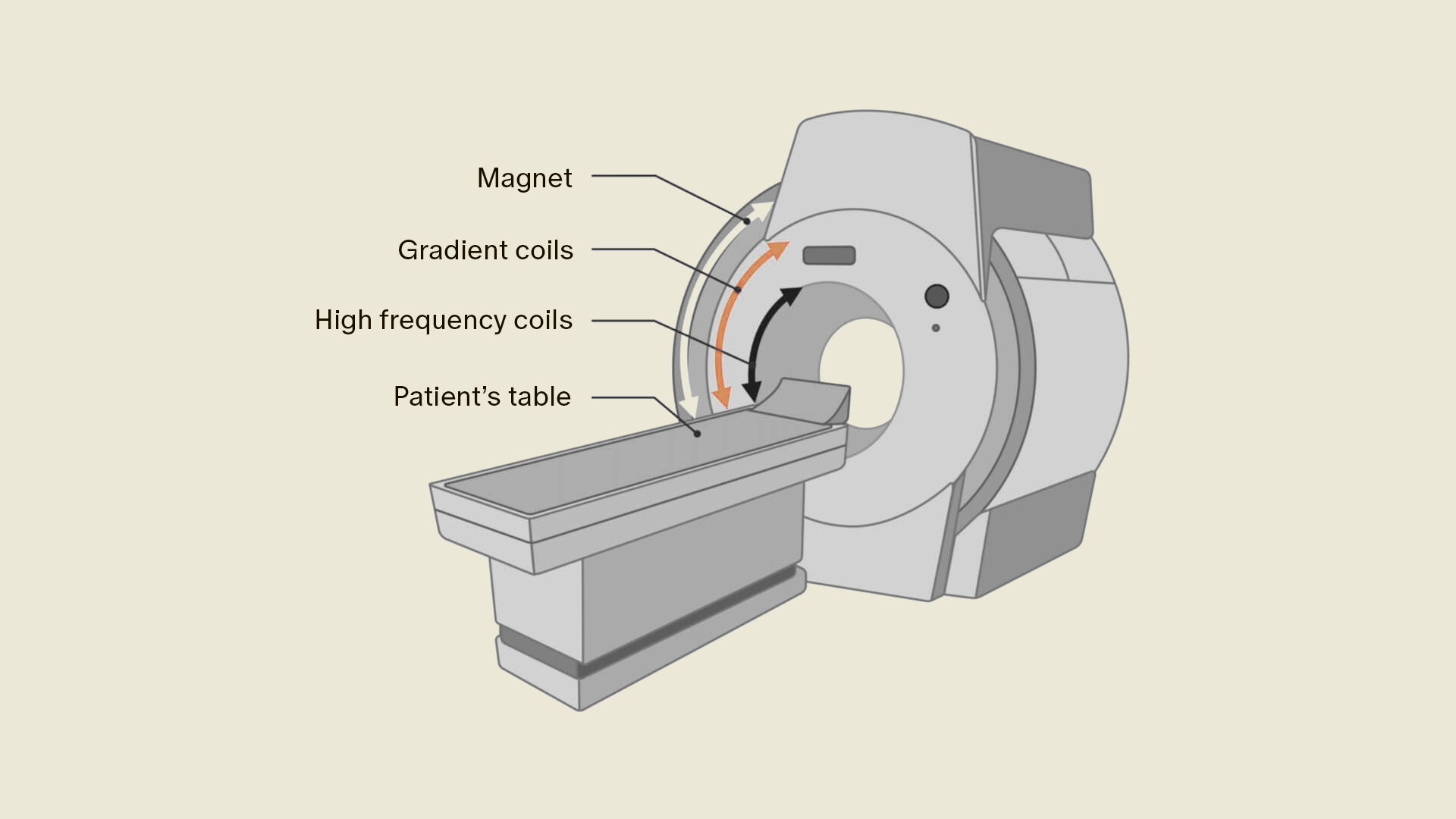
In an MRI, images of the body's interior are created by utilizing the hydrogen atoms within. These hydrogen atoms have protons that usually move in random directions. When a strong magnetic field is applied, these protons align in a specific direction. Radio waves are then sent to them, causing the protons to move. The device measures this movement and uses it to produce images of the body's interior.
To create accurate images, various magnetic fields acting in different directions are required. These additional magnetic fields come from special magnetic coils within the machine. These coils must constantly turn on and off and adjust themselves to capture images from different areas of the body. The frequent switching of these magnetic coils is the reason for the typical knocking noises heard during an MRI scan.
To reduce noise levels, you will be provided with hearing protection (headphones or earplugs) to dampen the sounds. At Aeon, you can even listen to music during your MRI scan. Throughout the entire scan, you can communicate with our medical experts to ensure you feel comfortable.
In most cases, you can resume your normal activities immediately after the scan. At Aeon, the images are analyzed by a Swiss radiologist, and you will receive the results within 72 hours, which you can discuss with your doctor.
CT
A CT scan is generally simple and painless, as long as no contrast agent needs to be injected. It usually takes less time than an MRI, typically between 5-15 minutes.
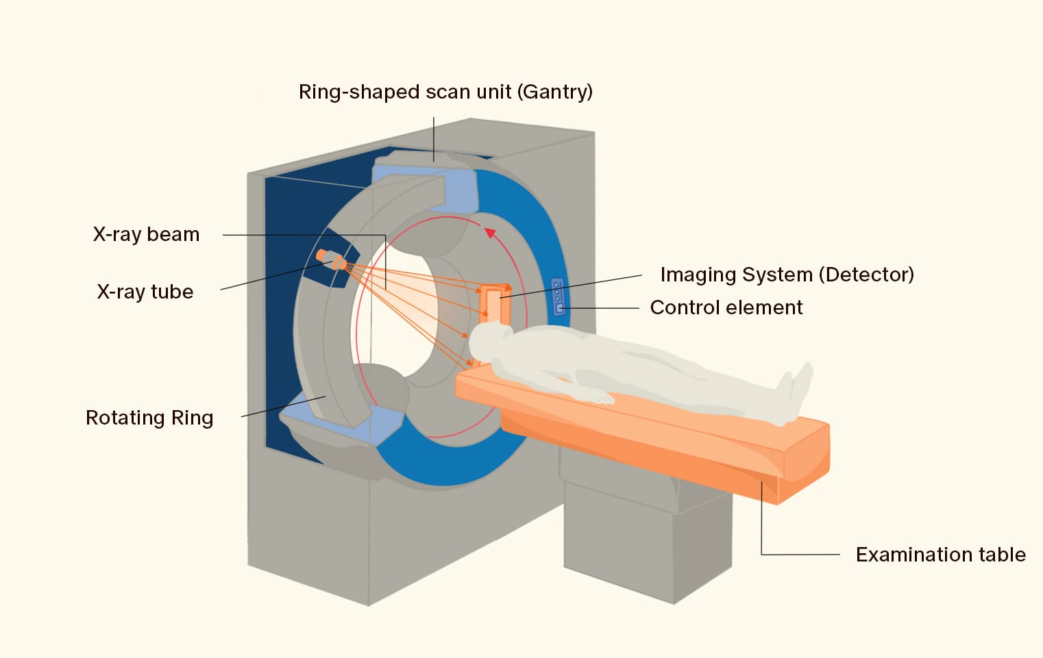
Unlike an MRI, metal parts do not pose a problem for a CT scan since CT scans do not use magnetic fields but instead rely on X-rays. These X-rays pass through your body and are captured by detectors on the opposite side. Both the X-ray tube and detectors rotate around the patient, taking numerous cross-sectional images of the body from different angles. A computer then assembles these into a detailed 3D image of your body’s interior.
After a CT scan, you can typically resume your normal activities immediately.
Radiation Exposure
Unlike CT scans and traditional X-rays, Magnetic Resonance Imaging (MRI) does not use ionizing radiation. As there is no risk of radiation exposure (e.g., risk of cancer), MRI is the safer option, especially for repeated use, including for children and young adults.
Application Areas
A CT scan is particularly effective for imaging bone structures. It is commonly used to assess bone fractures, calcifications, or other bone-related conditions. Additionally, it provides detailed images of the lungs, making it useful for diagnosing pulmonary embolisms, pneumonia, and lung tumors.
On the other hand, an MRI offers better soft tissue differentiation, making it ideal for imaging the spinal cord, muscles, tendons, ligaments, internal organs, and blood vessels. This is why it is highly effective for detecting changes in soft tissue, especially inflammation and tumors.
If you'd like to learn more about when an MRI is recommended, check out our detailed article here.
Contrast Agents
When contrast agents are used, different substances are employed for CT and MRI due to the differences in how the scans work.
For CT scans, iodine-based contrast agents are typically used because iodine absorbs X-rays more effectively.
For an MRI, the contrast agent usually contains gadolinium, which interacts with magnetic fields to enhance image contrast.
The use of contrast agents in both CT and MRI must be carefully considered by the attending physician. In some cases, they are essential for highlighting specific tissues or structures and providing more accurate diagnoses. However, contrast agents can also cause side effects (such as nausea and headaches) and are generally not used for pregnant patients or those with known allergies to the agent or kidney dysfunction.
For more information about the side effects of contrast agents, click here.
At Aeon, we avoid using contrast agents to eliminate the risk of side effects.
Conclusion
Whether you should undergo a CT or MRI scan for preventive care depends on several factors. A CT scan is usually faster, but an MRI allows for an examination without radiation exposure. Iodine-based contrast agents used in CT scans are more taxing on the kidneys than gadolinium in MRI scans. However, CT scans are often better suited for patients with metal implants.
Ultimately, the decision depends on what needs to be examined. As a general rule of thumb: for bones and lungs, choose CT; for muscles, soft tissues, the spine, internal organs (except lungs), and blood vessels, choose MRI.
You can book a full-body MRI check-up with us here.


