Discover how MRI provides detailed, radiation-free images for disease diagnosis and monitoring.
Magnetic resonance imaging (MRI) of a living human was first performed in 1977. Since then, MRI imaging has continuously evolved and is increasingly used in various fields. An MRI is particularly well-suited for depicting soft tissues such as the brain, spinal cord, and internal organs, as well as ligaments, cartilage, and certain bony structures. MRI is used for preventive screenings, initial diagnoses, and monitoring the progression of many diseases.
In this article, we aim to provide you with an overview of MRI, when it is used, the different types of MRI, and how it differs from other imaging methods like X-ray or computed tomography (CT).
Key Facts
- MRI (Magnetic Resonance Imaging): Provides detailed images of nearly all internal structures without ionizing radiation.
- Soft Tissue Imaging: Ideal for imaging soft tissues like the brain, spinal cord, internal organs, ligaments, and cartilage.
- T1 and T2 Imaging: T1-weighted images show fluids as dark and fat as bright, while T2-weighted images show fluids as bright.
- Multidimensional Images: MRI can produce images from various planes, allowing for detailed visualization.
- Contrast Agents: Enhance visibility of certain structures but pose risks for those with kidney issues and allergies.
- Advantages over CT: Better soft tissue contrast, no radiation, and the ability to visualize blood vessels without contrast agents.
- Safety Considerations: Suitable for most, but caution needed for those with implants and severe claustrophobia.
- Costs in Switzerland: Range from CHF 1,000 to 4,500, depending on scan complexity and use of contrast agents.
What is an MRI?
Magnetic Resonance Imaging (MRI), also known as magnetic resonance tomography (MRT) or nuclear magnetic resonance imaging, is a non-invasive imaging technique that provides detailed images of almost all internal structures of the human body. MRI machines create cross-sectional images of the body using a strong magnetic field and radiofrequency (RF) pulses. Unlike X-ray and CT, there is no radiation exposure during an MRI exam.
How does an MRI work?
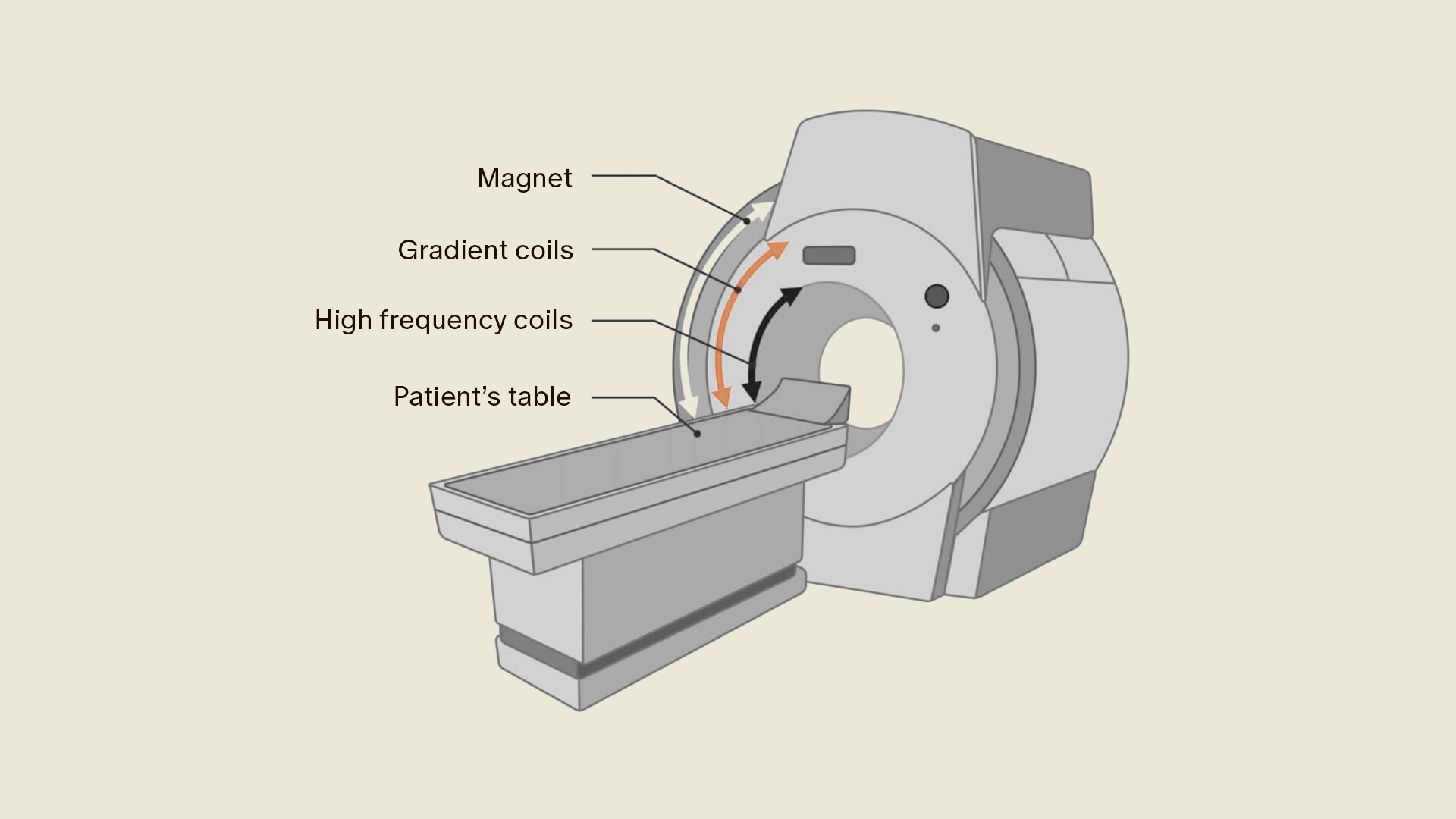
Humans are composed of 70% water, which includes hydrogen protons. These hydrogen protons, present in most body tissues, align in an MRI scanner along a strong magnetic field and are stimulated by an RF pulse. When the RF pulse is turned off, the hydrogen protons return to their original state. This is referred to as relaxation. Based on relaxation, different tissue types can be distinguished. Gradient coils can slightly alter the magnetic field, allowing different planes to be selected.
To obtain an MRI image, a body layer must be stimulated and measured multiple times. The image contrast is determined by T1 weighting and T2 weighting. Below, we explain what T1 and T2 mean and which regions are best depicted by each imaging method.
What does an MRI show?
An MRI scan can examine nearly all body regions, with internal organs, the musculoskeletal system, and blood vessels being particularly well depicted. One major advantage of MRI over other imaging methods is its excellent soft tissue contrast. This is due to the presence of hydrogen protons in many tissues, allowing MRI to provide detailed imaging of different tissue types.
Different tissue types have different relaxation times (T1 and T2), meaning they can be depicted differently.
By altering acquisition parameters, such as the repetition time (TR) or echo time (TE), different weightings can be achieved. Depending on the weighting, the various tissues are displayed in their characteristic signal intensity (gray values). Terms used include hyperintense (signal-rich, bright) and hypointense (signal-poor, dark).
Furthermore, MRI examinations can produce images in various planes (sagittal, axial, coronal) and slice thicknesses, enabling detailed visualization from different angles and 3D images.
T1-weighted Imaging
The T1 relaxation time, also known as longitudinal relaxation time, is the time it takes for the magnetization of a proton to be restored in the direction of the static magnetic field after being altered by an RF pulse. In T1-weighted images, fluids are depicted as dark, while fat (e.g., visceral fat, bone marrow) appears bright.
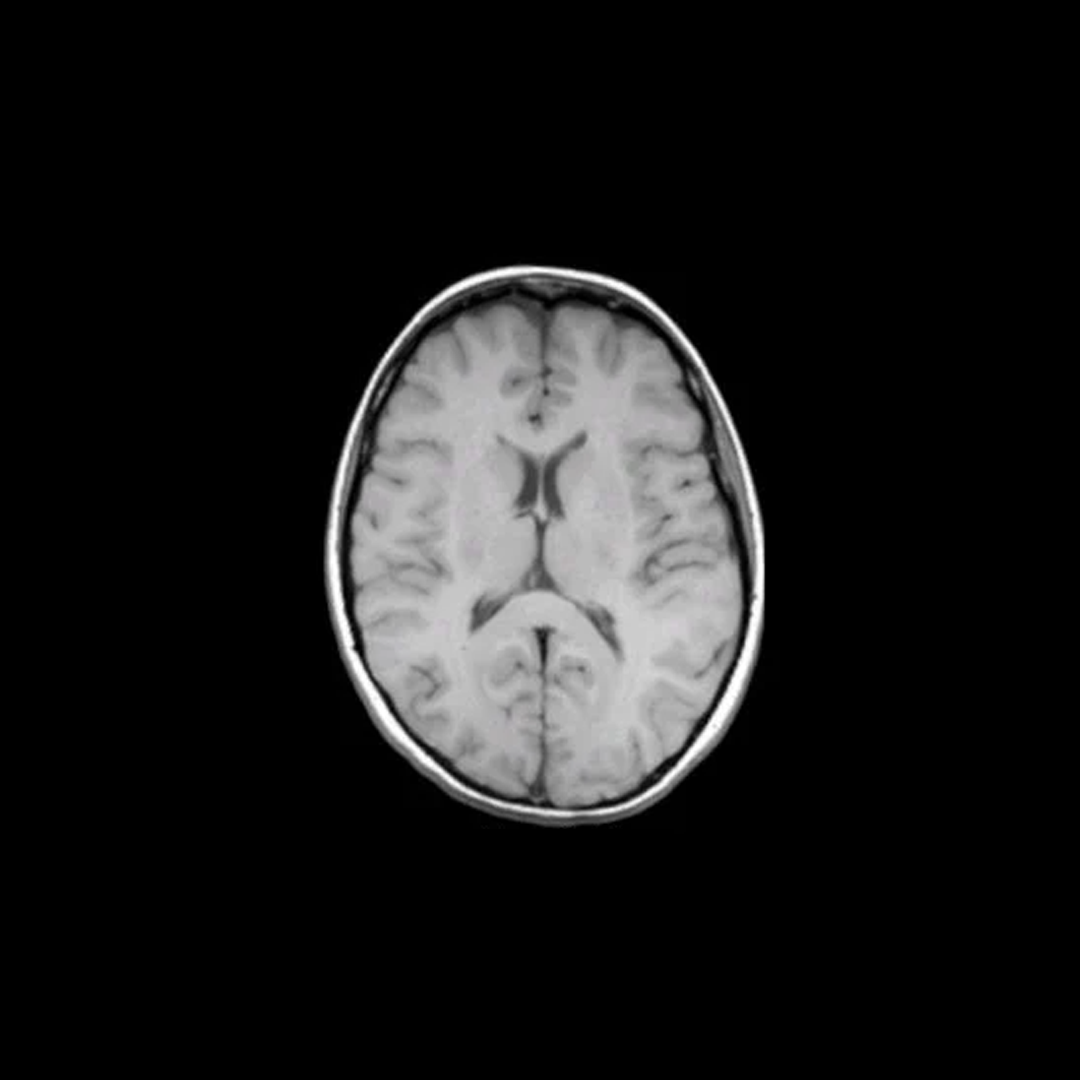
Graphic 1: Axial skull MRI in T1 weighting
T2-weighted Imaging
The T2 relaxation time is the period it takes for protons to return to their original state within the transverse plane after an RF pulse. Fluids, such as bile or cerebrospinal fluid (liquor), appear bright on T2-weighted images due to their long T2 relaxation times.
Due to their increased fluid content, most diseased tissues appear more signal-intensive and thus brighter than healthy tissues.
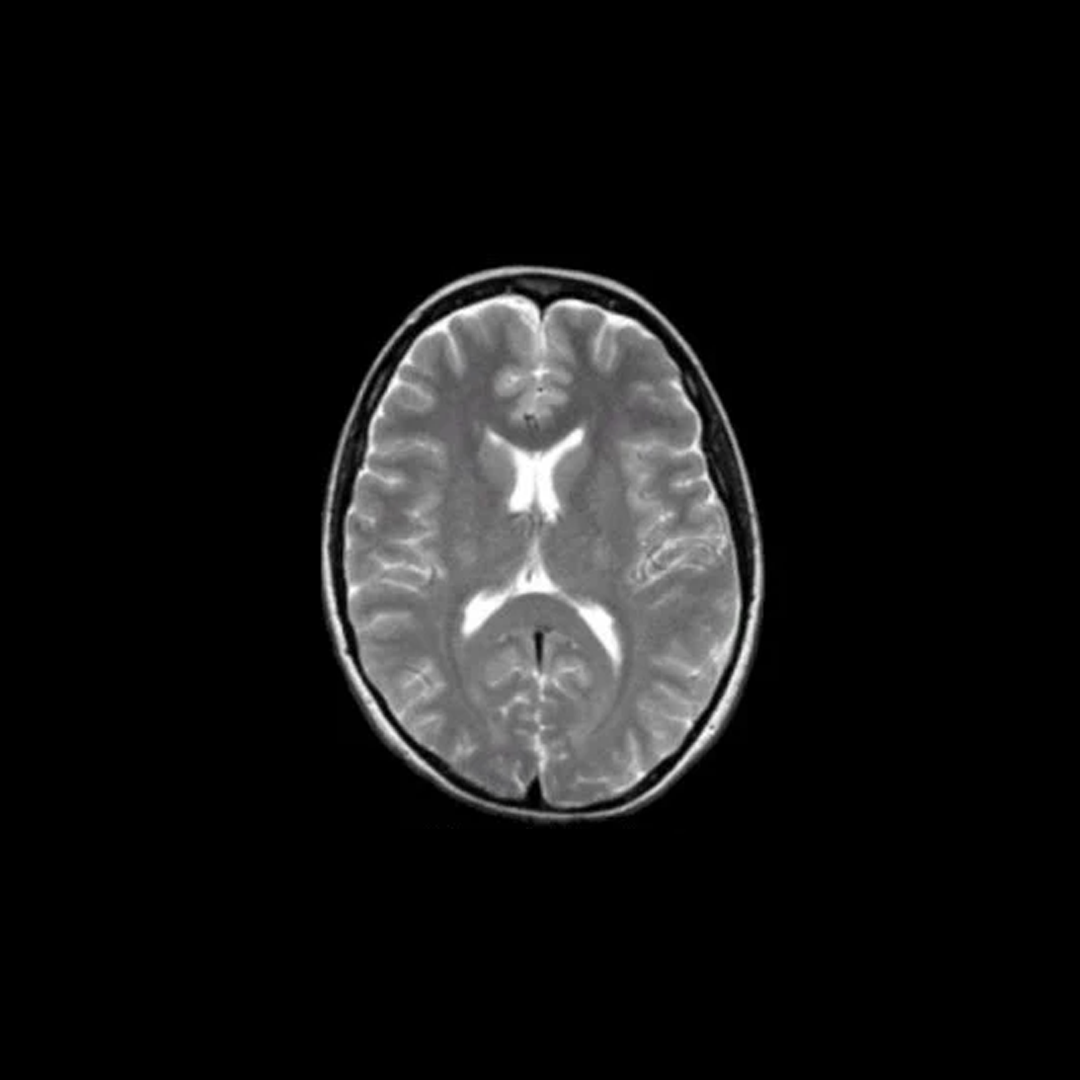
Graphic 2: Axial skull MRI in T2 weighting
Proton Density (PD)-weighted Imaging
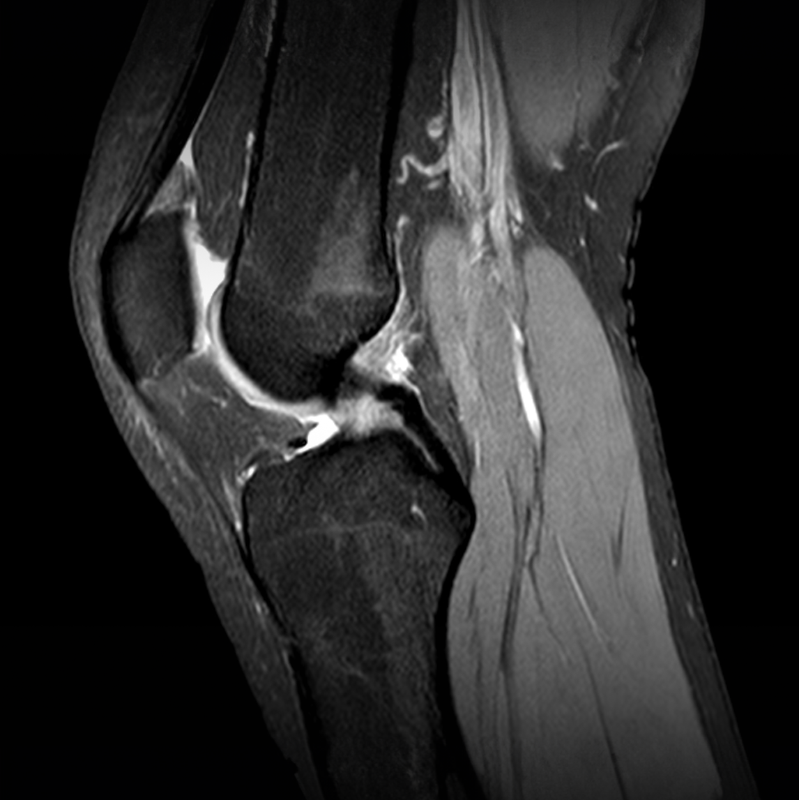
PD-weighted images in MRI utilize the number of protons in the examined tissue. Proton density contrast differentiates tissues in the body. Compared to T1 and T2-weighted images, they sometimes provide additional diagnostic information and complement these. PD-weighted sequences are particularly used in musculoskeletal imaging.
Diffusion-weighted Imaging
Diffusion-weighted imaging (DWI) measures and displays the movement of water molecules in various tissues of the body. These diffusion-weighted images can provide information about tissue structure and integrity, as the diffusion movement of hydrogen protons in tissues can be influenced by various pathological processes.
DWI is frequently used for the diagnosis and assessment of diseases such as strokes and their differentiation, inflammations, and tumor diseases, as it can detect changes in tissue diffusion early on.
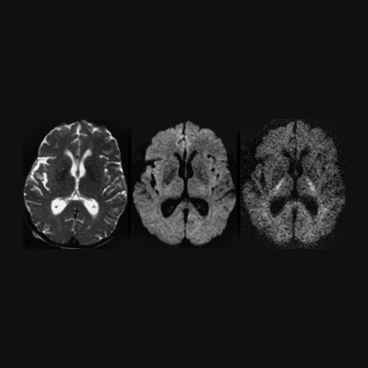
Graphic 4: Axial diffusion-weighted skull MRI ~~~~
MR Angiography (MRA)
MRI is excellent for depicting vessels, both within the skull and other body sections. MRA can be performed both with and without the administration of contrast agent.
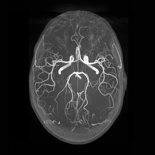
Graphic 5: MR Angiography of the cerebral arteries: axial maximum intensity projection of a Time-of-Flight MRA; the arteries are brightly depicted.
When and why should an MRI be conducted?
There are a variety of diseases that can be diagnosed using an MRI examination. These include tumor diseases, inflammatory processes, degenerative changes of the bones and spine, vascular diseases, as well as diseases of the central and peripheral nervous system. In this article, we provide you with an overview of the most important MRI examinations and the diseases or changes that can be detected therein.
Who is not suitable for an MRI examination?
There are certain groups of people who may not be suitable for an MRI examination or for whom special precautions may be necessary. Before an MRI examination is conducted, the medical staff conducts a thorough medical history to identify potential risk factors and ensure that the examination can be carried out safely and effectively. Below, we provide you with an overview:
Individuals with implanted medical devices
Some electronic devices may be affected by the strong magnetic fields of MRI machines. This includes:
- Pacemakers
- Neurostimulators
- Mechanical heart valves
- Insulin pumps
- Cochlear implants
In such cases, special safety precautions must be taken before the MRI examination. In some cases, an MRI examination is not possible, and an alternative imaging method must therefore be used.
Individuals with metal implants or foreign bodies
Metallic implants, prostheses, stents, clips, screws, copper coil, or other metal objects (e.g., metal shrapnel) in the body can also cause problems during an MRI examination. Depending on the type and location of the metal, these objects can disrupt the image or lead to injuries. It is important to identify and review all metal objects in the body before the examination.
Kidney diseases (only relevant with contrast agent administration)
People with severe kidney dysfunction or renal insufficiency may not be able to receive an MRI with gadolinium-containing contrast agent, as this contrast agent is excreted through the kidneys. At aeon, we do not use contrast agents.
Claustrophobia Also, individuals with claustrophobia may have difficulty remaining calm during the MRI examination. At aeon, we have MRI machines with a larger tube diameter to ensure the comfort of our customers. We offer h
Pregnant women
Although there is no clear evidence of a link between MRI and subsequent biological effects on the fetus, an MRI during pregnancy is generally avoided unless there is an urgent medical need. In such cases, the potential risks to the mother and unborn child must be carefully weighed against the benefits of the examination.
What is the MRI examination process?
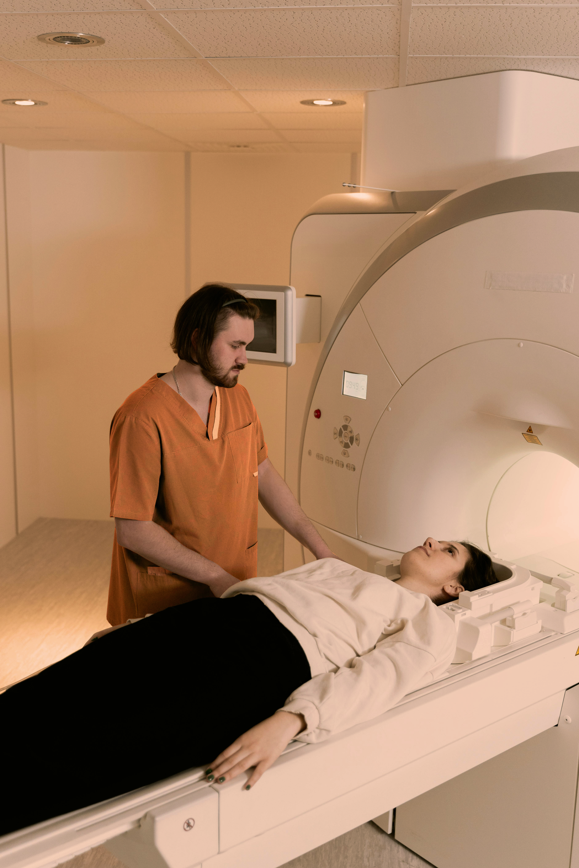
An MRI examination is a straightforward and pain-free process that typically takes 15-60 minutes, depending on the body region being examined. No radiation is used. You will be positioned lying down on a special table that is moved into the opening of the MRI machine.
If you feel uncomfortable or have questions, you can communicate this during the examination. It is important to us to make your MRI examination as comfortable as possible.
The process is as follows:
Preparation
Before the examination begins, you will be asked to remove jewelry, piercings, metal objects, and clothing with metal parts. The radiology staff should also be informed about any fixed metal objects in the body (e.g., spiral or pacemaker, see above).
Positioning
You will be placed on a movable table, which is then pushed into the MRI scanner. It is important to remain still throughout the examination, as movement can affect image quality.
Contrast agent (optional)
In some cases, it is decided by the treating physician whether a contrast agent is used to make certain structures or tissues more visible. This contrast agent is usually administered through an intravenous (IV) injection, either before or during the examination. At aeon, we do not use contrast agents.
Conducting the examination
During the MRI examination, you lie in a tube within the MRI scanner. While the images are being taken, you may hear loud knocking sounds caused by the switching on and off of the gradient coils. You will receive hearing protection (headphones, earplugs) to dampen these noises. At aeon, we use modern scanners with a large tube diameter to increase comfort. With us, you can also listen to music and watch videos during your MRI examination, and you will be accompanied by our professional staff throughout your examination.
Completion
Once the images have been taken and the examination is complete, you will be moved out of the MRI scanner. In most cases, you can resume your normal activities immediately after the examination, unless special instructions have been given or you have received a sedative beforehand.
The entire MRI examination typically lasts between 15 and 60 minutes, depending on the region being examined and the specific requirements of the physician. The images are evaluated by a radiologist, and the results will be communicated to you shortly after the examination.
What are the advantages of an MRI compared to other examination methods?
- No ionizing radiation: Unlike computed tomography (CT) and X-ray examinations, MRI does not use ionizing radiation. This means it is safer for the patient and carries no risks associated with radiation exposure (e.g., skin damage, cancer development).
- Better soft tissue contrast: MRI offers significantly superior soft tissue contrast compared to CT and X-ray examinations. This allows doctors to obtain more detailed images of organs, tissues, muscles, tendons, and other soft tissue structures.
- Multidimensional images: MRI can produce images from various planes, thus enabling a detailed and comprehensive assessment of the examined body area. This is particularly beneficial for complex anatomical structures and pathological processes.
- Contrast agents without iodine: In MRI, contrast agents that do not contain iodine can be used, which is beneficial for people with allergies or kidney problems, as MRI contrast agents have a lower risk profile compared to iodine-containing contrast agents.
- Vessel depiction without contrast agents: Advanced MRI techniques, such as MRI angiography, can visualize blood vessels without the use of contrast agents, which is useful for diagnosing vascular diseases such as aneurysms or stenoses.
- Functional imaging: Functional MRI (fMRI) is a special application of MRI that measures the activity of specific brain regions during specific tasks. This is an important tool in neuroscience and is frequently used to study brain functions and diagnose neurological diseases.
Overall, MRI is a safe, non-invasive, and extremely versatile imaging method with excellent soft tissue depiction and various applications in the diagnosis and monitoring of diseases and conditions.
What is the difference between a CT and MRI?
Both a CT scanner and an MRI scanner produce detailed images using very different techniques. The main difference is that an MRI machine works with RF waves (radio waves) and a strong magnetic field, while a CT scanner uses X-rays. Both types of scans are considered relatively low-risk and safe when used appropriately.
CT is particularly well-suited for depicting bone structures and is often the preferred modality for assessing fractures, other bony pathologies, and calcifications. It provides detailed images of the lungs and is frequently used to diagnose pulmonary embolisms, pneumonia, and lung tumors. The high air content of the lungs can complicate or distort imaging with MRI. Furthermore, CT can be performed very quickly and is therefore well-suited for emergency situations.
In contrast, MRI provides better soft tissue differentiation and is ideal for depicting soft tissue structures such as the brain, spinal cord, muscles, tendons, ligaments, internal organs, and blood vessels. It is particularly helpful in diagnosing tumor diseases, inflammatory processes, diseases of the central and peripheral nervous systems, vascular diseases, degenerative changes, diseases of the spine, and joint injuries. MRI does not use ionizing radiation, making it a safer option for repeated applications, especially in children and young adults.
Contrast agents - yes or no?
The use of contrast agents in MRI examinations is a decision that must be carefully weighed. In some cases, the use of contrast agents may be sensible to better depict certain tissues or structures and to more accurately diagnose specific diseases. Contrast agents can be used, for example, to visualize blood vessels, tumors, inflammations, or other lesions with increased blood flow.
However, there are also situations in which it is better to forego contrast agents. This can have various reasons, including:
- Kidney dysfunction: People with impaired kidney function may be at increased risk for permanent kidney damage and systemic involvement (NSF: Nephrogenic Systemic Fibrosis) if they receive contrast agents, as these are excreted through the kidneys. In such cases, it is important to carefully weigh the risks and benefits of administering contrast agents and possibly choose alternative examination methods.
- Allergic reactions: Some individuals may be allergic to contrast agents, leading to undesirable side effects such as skin rashes, itching, breathing difficulties, or rare severe reactions. Individuals with known allergies should inform the medical staff before administering contrast agents.
- Pregnancy: During pregnancy, it is usually advisable to avoid the use of contrast agents to minimize the risk to the fetus. In urgent cases, however, a careful risk-benefit assessment may be required.
The administration of contrast agents can lead to side effects. These include:
- Pain at the injection site
- Nausea
- Itching
- Skin rash
- Headaches and dizziness
- Taste disturbances
- Visual disturbances
- In patients with severe renal insufficiency, serious side effects may occur in rare cases in the frame of Nephrogenic Systemic Fibrosis (NSF).<
At aeon, we do not use any contrast agents.
Are there other side effects of an MRI?
Generally, magnetic resonance imaging (MRI) is a safe and non-invasive imaging examination method that does not use ionizing radiation. Most people experience no side effects during or after an MRI examination.
However, in rare cases, some side effects may occur, including:
- Claustrophobia: Some patients may experience a feeling of constriction or anxiety during the examination, especially if they suffer from claustrophobia (fear of enclosed spaces).
- Heat sensation: Some patients may experience a temporary heat sensation during the injection of the contrast agent, which spreads through the body. This is usually harmless and disappears quickly.
- Metallic objects: Metallic implants or foreign bodies in the body may be heated during the MRI examination or, in very rare cases, move, causing discomfort or even injuries. Therefore, it is important to inform the medical staff of all known implants or foreign bodies in the body.
How much does an MRI cost in Switzerland?
An MRI costs approximately CHF 1,000 to CHF 4,500 in Switzerland, depending on the body region and duration of the scan. Additionally, the costs vary depending on whether the examination is conducted with or without contrast agents. If multiple structures are examined (whole-body scan), the MRI may take longer and the costs may be higher.
In some cases, an MRI examination is covered by health insurance, especially if it involves a partial scan of specific body regions. Whole-body scans are usually not covered and are not offered by most facilities. Additionally, radiologists are often not familiar with the evaluation of whole-body scans, as they typically specialize in single body regions and organs. At Aeon, we have specially trained doctors who, supported by highly specialized AI imaging technology, expertly and detailedly interpret your scan.
Conclusion
Magnetic resonance imaging (MRI) is a non-invasive imaging technique that provides detailed images of almost all internal structures of the human body without using ionizing radiation. MRI offers superior soft tissue depiction and allows doctors to accurately differentiate between various tissue types and pathological processes.
MRI can be used for various purposes, including the examination of the head, chest, spine, bones and muscles, joints, blood vessels, and heart, as well as the internal organs of the abdominal cavity. It offers a safe and versatile imaging method with multiple advantages over other imaging methods such as computed tomography (CT) and conventional X-ray.
Although most people experience no side effects during or after an MRI examination, there are some rare risks and contraindications that must be considered, especially in individuals with implanted medical devices, metal implants, certain diseases, or during pregnancy. It is important to conduct a thorough medical history before an MRI examination to identify potential risk factors and ensure that the examination can be carried out safely and effectively.


