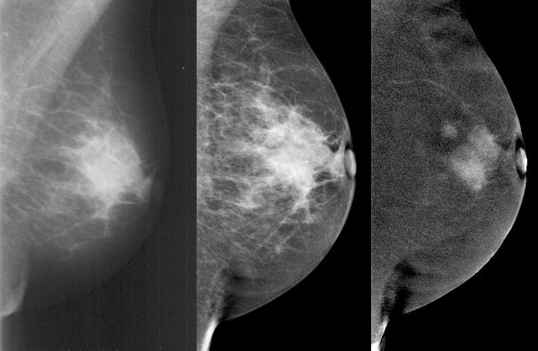
Mammography: X-ray examination for early detection of breast cancer. Learn all about the mammography procedure, screening programs, pros and cons, and available alternatives.
With more than 2.3 million cases per year, breast cancer is the most common cancer among adults. In 95% of countries worldwide, breast cancer is the leading or second leading cause of cancer-related deaths among women.
In Switzerland, breast cancer is also the most common cancer among women. Early detection plays a crucial role as it significantly improves survival rates. Studies have shown that women who participated in mammography screening had a 41% lower risk of dying from breast cancer within 10 years.
In this article, you will learn everything important about mammography, its benefits and risks, as well as recommendations for women in Switzerland.
What is a Mammography?
A mammography is an X-ray examination of the breast used for diagnosing or early detection of breast cancer (mammary carcinoma) and other breast abnormalities. Mammography images can reveal changes in breast tissue, and additional tests such as an ultrasound or MRI can be conducted if necessary.
Mammography is the most common method for the early detection of breast cancer. It allows for the identification of small tumors or changes in the breast, often in the early stages when they are still treatable.
When is a Mammography Performed?
Mammography is not only conducted as part of a screening program but can also be used earlier when abnormalities are found during an examination, in cases of pain, or if there is a family history of breast cancer:
- Positive palpation findings: If palpable changes in the breast are detected, a radiologist or gynecologist may perform an ultrasound (mammosonography) or mammography. Tissue samples are taken under visual guidance and examined by a pathologist.
- Bloody or serous discharge from the nipple: Discharge from the nipple can be a sign of breast cancer. According to one study, about 9% of women over 50 who experienced nipple discharge were diagnosed with breast cancer or a pre-cancerous condition.
- Breast asymmetry: If there is sudden breast asymmetry with no clear findings from palpation, mammography may be recommended. A study from the University of Liverpool found that women with larger asymmetry had a higher risk of developing breast cancer. For every 100-milliliter volume difference, the risk increased by 1.5 times.
- Screening program: In Switzerland, 13 cantons offer a breast cancer screening program, where women aged 50-74 can have a mammography every two years.
- Hereditary predisposition: If a mother or sister has had breast cancer, the risk of developing breast cancer doubles. According to the Cancer League, 5-10% of women with breast cancer have a genetic predisposition. Familial breast cancer is often linked to inherited gene mutations, commonly in the BRCA1 and BRCA2 genes. These mutations can be detected through a BRCA test.
How Does a Mammography Work?
- Registration: Women aged 50 to 74 receive an invitation for mammography and can schedule an appointment with a participating radiologist or breast center.
- Preparation: Before the examination, the patient is informed about the procedure, and any questions are answered. A health questionnaire (anamnesis) is completed, and any jewelry near the breast area is removed. Avoid using deodorant or creams on the breast area as they can affect the quality of the X-ray images.
- Positioning: Mammography is performed standing. The woman stands in front of the X-ray machine, and a radiologist or radiographer ensures the correct posture.
- Compression: Each breast is compressed between two plates to improve image quality and reduce radiation exposure.
- X-ray image: Two X-ray images of each breast are taken from different angles (frontal and lateral). The actual image capture takes only a few seconds, and the entire process takes about 10-15 minutes.
- Evaluation: The images are evaluated independently by two specialists, and results are typically communicated within two weeks via mail or phone. If the results are abnormal, further tests like an ultrasound, biopsy, or MRI may be recommended.
Is Mammography Painful?
The compression of the breast can be uncomfortable or even painful, depending on the woman and factors such as breast tissue density and the phase of the menstrual cycle. It is recommended that women with menstrual cycles undergo the exam during the first half of their cycle when breast tissue is less tender.
What Can Be Seen in Mammography Images?
Mammography can detect various abnormalities, including benign tumors (fibroadenomas), cysts, lumps, or small calcium deposits (microcalcifications), which may indicate cancer or a precancerous condition.

What Are the Advantages of Mammography?
- Reduced Mortality: Regular mammography screening significantly reduces breast cancer mortality rates. Annual screenings for women between 40 and 84 can lower mortality by 40% compared to no screening.
- Early Detection: Mammography detects tumors earlier, when they are smaller and have not spread to the lymph nodes, leading to a much higher survival rate (99% for localized cancers) and less aggressive treatment options.
- Less Morbidity from Treatment: Early detection allows for less invasive treatments, such as lumpectomy (breast-conserving surgery) instead of mastectomy (removal of the breast gland), reducing the risk of complications like pain and lymphedema.
What Are the Disadvantages of Mammography?
- Radiation Exposure: While mammograms can prevent many deaths from breast cancer, the radiation exposure can lead to new breast cancer cases. A less frequent screening (every two years starting at 50) can significantly reduce this risk.
- False Positives: Up to 20% of women between ages 50 and 69 experience a false positive result, with 3% undergoing unnecessary biopsies.
- False Negatives: Up to 35% of breast cancer cases may be missed by mammography.
- Overdiagnosis: Some cancers detected by mammography may never cause symptoms, leading to overdiagnosis. One meta-analysis found 12.6% of women over 40 who had mammograms were overdiagnosed.
- Pain: 77% of women reported pain during mammography, primarily due to the strong compression of the breast tissue.
How Much Does a Mammography Cost in Switzerland?
The cost of a mammography is 90% covered by health insurance, with a co-pay of 10%. Depending on the canton, this amounts to between CHF 17.25 and CHF 182.80. For cantons without a screening program, costs are not covered by insurance.
Are There Alternatives to Mammography?
While mammography is effective, it is not without limitations. Alternative methods include breast MRI and ultrasound, which may be more suitable for younger women or women with dense breast tissue.
Since many women find mammography painful, we present the following other breast cancer screening options, which are currently not covered by insurance:
| Examination | Advantages | Disadvantages | Detection Rate |
|---|---|---|---|
| Breast MRI |
|
|
90–95 % |
| Breast Ultrasound (Sonography) |
|
|
Approx. 50 % |
| Breast Tomosynthesis (DBT) |
|
|
In combination, up to 48% higher than standard mammography |
Conclusion
Mammography plays a crucial role in the early detection of breast cancer, the most common cancer among women worldwide. Regular screenings can detect tumors at an early stage, significantly improving the chances of recovery. In Switzerland, several cantons already offer screening programs for women aged 50 to 74.
While mammography is effective, it is not without risks. Besides radiation exposure, there is the possibility of false-positive or false-negative results and overdiagnosis. Additionally, the compression during the examination can be painful.
Breast MRI, with its high detection rate, suitability for dense breast tissue, and pain-free procedure, offers a good alternative to mammography.
Would you like to learn more about the possibility of using MRI for early detection? Simply book a scan to gain valuable insights in your health.


